Spatial Proteomics Maps Tumor Ecosystems in Ovarian and Kidney Cancer
Studies in ovarian and kidney cancer show how spatial protein mapping and AI may reveal tumor features linked to recurrence, survival and treatment response.
Spatial maps of tumor cells may help explain why cancers with the same diagnosis can behave differently. In two studies discussed in a webinar featuring Bernd Bodenmiller, PhD, researchers used highly multiplexed protein imaging, molecular profiling and artificial intelligence to identify tumor patterns associated with recurrence, survival and treatment response. The findings offer a research framework for improving patient classification; they don’t establish a new clinical test or prove that changing treatment based on these patterns improves outcomes.
Why the tumor’s arrangement matters
A tumor contains more than cancer cells. Immune cells, connective tissue cells and other parts of the surrounding environment interact with cancer cells, and their locations can shape how a tumor develops or responds to treatment. Measuring which proteins are present while preserving information about where cells sit can therefore reveal features that a tissue sample’s overall molecular averages may miss.
Imaging Mass Cytometry, or IMC, is one way to do this. It can image dozens of proteins and transcripts in tissue at once, allowing researchers to describe cell types and their positions in a tumor sample. Adding other molecular data and computational analysis can help connect those local patterns with clinical outcomes. Still, a pattern associated with an outcome is a clue for research, not by itself evidence that the pattern causes that outcome.
Ovarian cancer: patterns linked to recurrence
One example covered in the presentation involved 323 epithelial ovarian cancers. Tumors with greater diversity in their cell phenotypes and spatial organization were associated with faster recurrence. The work also identified a tumor cell phenotype marked by TUBB3 and pRB proteins that was associated with recurrence and a poorer response to standard treatment.
The researchers investigated these cells with spatial transcriptomics and drug screening in 3D sphere models and patient samples. The reported results suggest that the cells resist standard agents while remaining sensitive to certain inhibitors. That contrast may help researchers identify treatment vulnerabilities worth studying further. It does not show that these inhibitors are effective or appropriate for patients with this phenotype; the account describes laboratory and sample-based findings, not a clinical trial demonstrating patient benefit.
The distinction matters for interpreting treatment-resistance research. Finding a candidate vulnerability in cells or model systems can guide the next experiment, but clinical studies are needed to establish whether a treatment works in people, which patients may benefit, and what harms might occur. The source does not provide details such as trial results, treatment doses or a validated test for identifying these cells in routine care.
Kidney cancer: survival ecotypes from tissue images
A second study examined 498 clear cell renal cell carcinomas. Using AI, the researchers identified three survival ecotypes: groups defined by patterns in the tumor ecosystem that were associated with different survival outcomes. The groups also had distinct genomic and metabolic features, and each was predictive of treatment outcome.
Deep learning was used to predict these ecotypes directly from routine hematoxylin and eosin, or H&E, tissue slides. The findings were validated across five cohorts totaling more than 2,500 patients. Validation across several cohorts makes the result more persuasive than a pattern observed in just one patient group, though it still doesn’t establish that the classification can guide treatment in everyday practice. The source does not report the ecotypes’ performance measures or describe a prospective study in which clinicians used them to select therapy.
The comparison with basic clinical information and molecular stratification models is also relevant: the ecotypes reportedly predicted survival better than those approaches. That result suggests that tissue architecture may contain information not captured by those other classifications. It doesn’t mean the image-based method replaces established clinical assessment, or that it will perform equally well across every patient population and care setting.
What this research may change
Together, the examples show how spatial protein imaging, other molecular measurements and AI can describe tumors in more detail than a single marker or tissue-wide average. The potential value is practical: a clearer map of cell populations and their relationships could help researchers distinguish tumors that look similar under current categories but have different outcomes or treatment responses.
The next questions are whether these patterns can be measured consistently, whether they hold up in further patient groups, and whether using them to guide decisions improves outcomes. For patients and families, the findings are not a reason to change treatment. Anyone wondering whether a tumor profiling result has implications for care can discuss the test’s evidence and limitations with their oncology team.
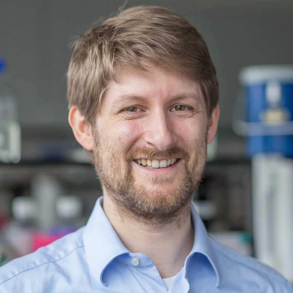
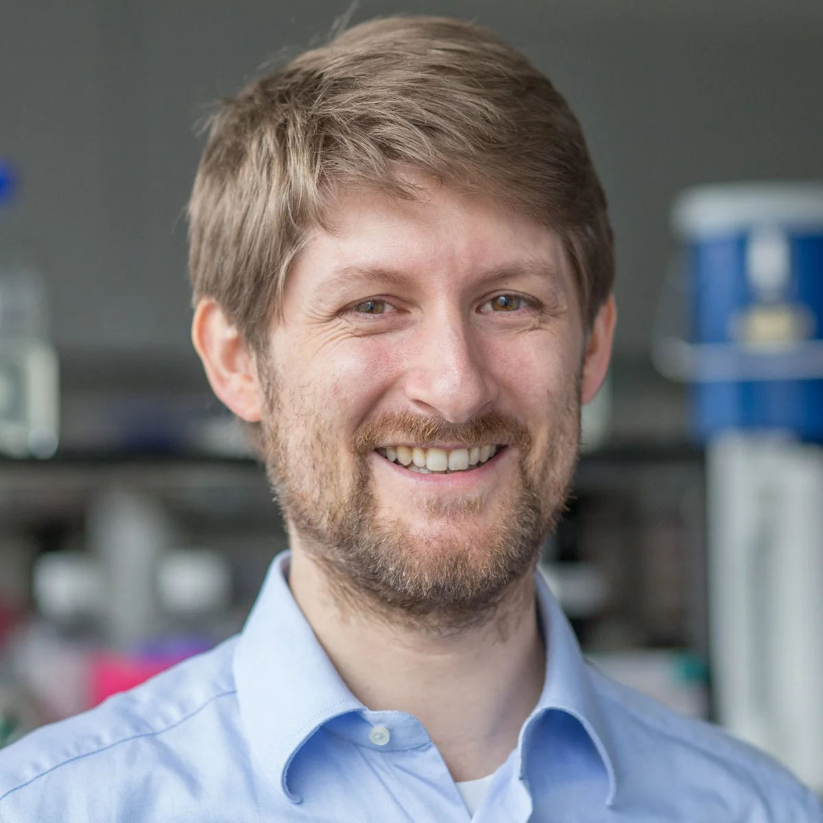
Bodenmiller, an associate professor at the University of Zürich and ETH Zürich, develops experimental and computational methods to study tumor ecosystems. His group pioneered IMC and the histoCAT software toolbox, which supports analysis of cells and their organization in tissue. The webinar is scheduled for Monday, November 16, 2026, at 08:00 PST, 11:00 EST and 16:00 GMT, with a live question-and-answer session after the presentation.
A live Q&A session will follow the presentation, giving attendees an opportunity to ask questions.
Produced with support from:


The post Spatial Proteomics for Precision Oncology appeared first on GEN - Genetic Engineering and Biotechnology News.