Gene Therapy Restores Some Light Sensitivity in Retinitis Pigmentosa Trial
In a 10-person trial, gene therapy paired with amber-light goggles improved some participants’ light sensitivity, but vision remained limited.
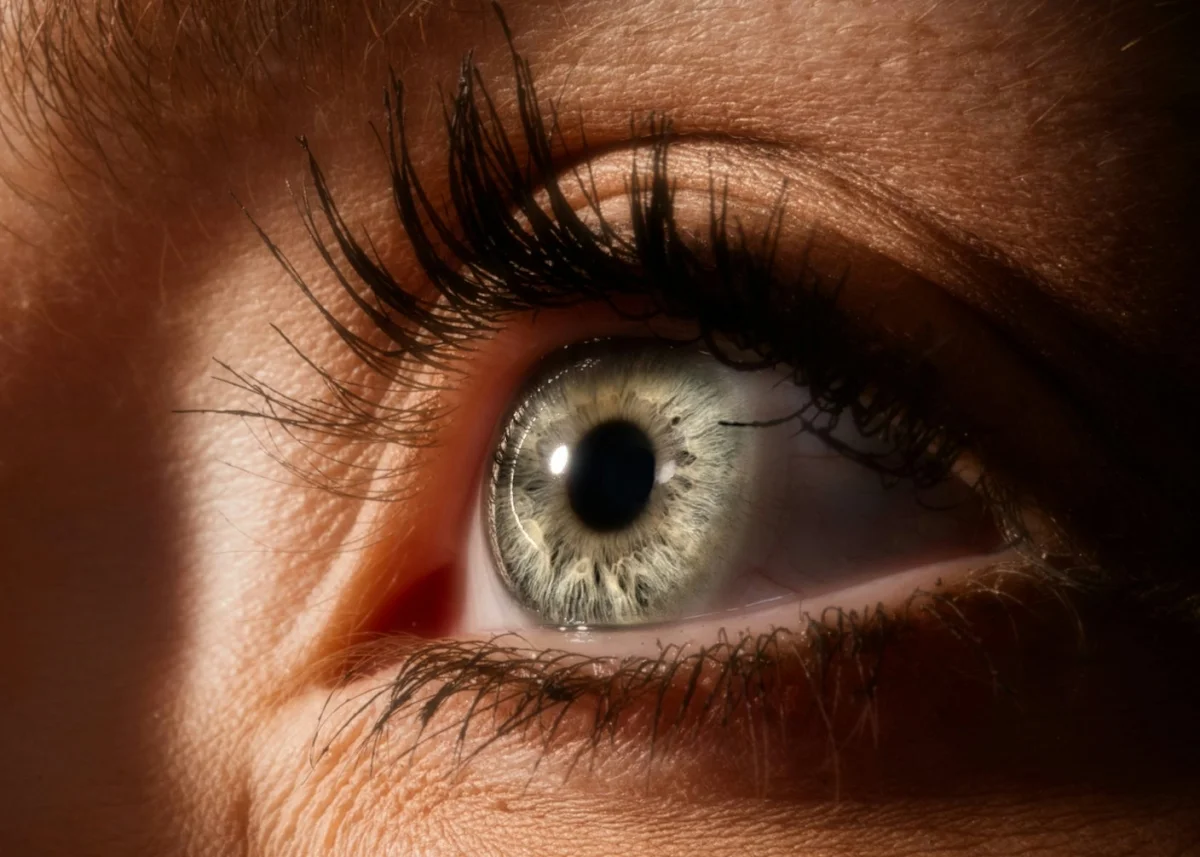
A small clinical trial suggests that gene therapy can give some people with advanced retinitis pigmentosa a limited ability to detect objects and light. The treatment didn’t restore reading or face recognition, and it requires specialized goggles. Its significance is narrower but meaningful: early gains seen in a single patient appear to have been repeated in a group, with improvements lasting for years among some participants.
The PIONEER study treated 10 people, ages 40 to 73, who were legally blind. Researchers injected genetic instructions for an algae-derived light-sensitive protein into each participant’s poorer-seeing eye. Seven received one of three increasing doses. The study’s main goal was to assess safety; tests of vision provided an early look at whether the approach might work.
After treatment, six participants had a clinically meaningful increase in light sensitivity, defined in the study as more than four times their baseline measurement. One participant’s reading rose by more than 62 times. Eight people went on to further vision tests; roughly half could locate and touch objects or identify the orientation of a bar. These results point to partial visual function, not sight comparable to that of a person with healthy vision.
Turning surviving cells into light sensors
Retinitis pigmentosa is a group of inherited conditions that gradually damage the retina’s light-sensing cells. More than 100 genetic variations have been linked to the disease. That diversity can make a treatment aimed at correcting one particular gene unsuitable for many patients.
The researchers instead used optogenetics: changing cells so they respond to light. The therapy delivered instructions for ChrimsonR, a protein originally found in algae, into retinal ganglion cells. These cells normally carry visual signals toward the brain; they don’t detect light themselves. Because they can persist after light-sensing cells have deteriorated, they may offer another route for sending visual information to the brain.
ChrimsonR responds to amber light. That feature allows the team’s goggles to activate the altered cells from outside the eye. A camera in the device scans the wearer’s surroundings and sends amber light pulses into the eye in response to changes across the scene. The system is designed to turn the environment into signals the engineered cells can detect.
This is a different strategy from earlier research that sought to make cone cells, which are among the retina’s light-sensing cells, responsive to light. In advanced retinitis pigmentosa, however, cones may already be too damaged to serve that role. Targeting the more resilient ganglion cells aims to work around that loss rather than repair the original sensors.
What participants could see
The visual information remained crude. Participants who responded could perform basic tasks such as finding a notebook or a small box and distinguishing changes in a rotating bar. They couldn’t read or recognize faces. Reports described blurry images, a narrow field of view and distorted-looking edges. The results suggest that the brain can make use of signals from the altered cells, but the detail available so far is limited.
Some participants’ gains continued over roughly five years. One person whose earlier experience drew attention in 2021 can now recognize and count objects, and can pick out white stripes on a crosswalk. That account is one example, not a measure of what every participant can expect.
Researchers also looked for signs that the brain was receiving useful visual input. In a test involving a tumbler, participants opened or closed their eyes while looking at it. The team found that vision-related electrical activity in the brain was more coherent when their eyes were open. This supports the possibility that the improved performance depended on visual signals, though it doesn’t establish how much useful sight the therapy can provide in daily life.
Safety and next steps
Most participants had temporary inflammation or increased pressure in the treated eye. The study reported no detectable effects throughout the body, and the retina’s structure remained unchanged. One participant had a blockage in a major eye artery that cleared within minutes after treatment with an eye drop. The report describes the injection as relatively safe in this small study, but 10 participants can’t reveal every possible risk or establish safety for broader use.
Earlier work helped build the rationale for the approach. Researchers showed that channelrhodopsin proteins could make nerve cells respond to light, and experiments in mice and human retina samples suggested that optogenetics might restore light sensitivity. The PIONEER findings add human trial evidence, while remaining an early result rather than proof of dependable functional vision.
Only some ganglion cells receive the therapy, which limits the amount of visual information they can carry. Researchers are exploring more light-sensitive versions of the protein and changes to the goggles, including eye tracking and software that processes scenes before stimulation. These are possible refinements; the trial hasn’t shown that they will produce sharper vision.
For people living with retinitis pigmentosa, this study offers a reason for cautious interest: a subset of participants gained measurable light sensitivity and basic object-detection ability, with some gains lasting years. The next question is whether larger studies can clarify who benefits, what daily tasks become easier, and how reliably the effect lasts.